Guangshou Ou’s group at School of Life Sciences at Tsinghua University published an article entitled "Modulation of inner junction proteins contributes to axoneme differentiation" in the journal Proceedings of the National Academy of Sciences of the United States of America (PNAS) on July 18, 2023, reporting on the mechanism and function of axoneme differentiation.
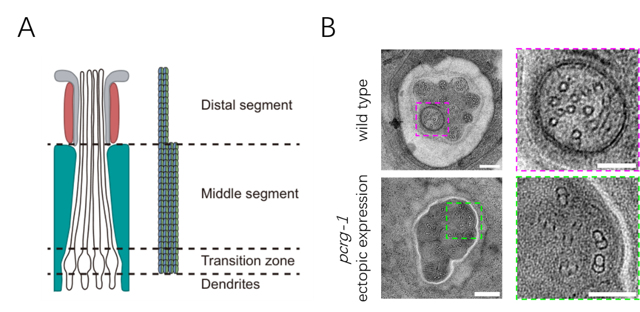
Cilia are organelles widely distributed on the surface of eukaryotic cells that enable cell movement and perception of external signals. The axoneme is a subcellular assembly based on microtubules that provides a scaffold for ciliary extension from the cell membrane. In sensory cilia of different species, the axoneme longitudinally differentiates into a middle segment with nine microtubule doublets and a distal segment with only microtubule singlets that extends from the A-tubules of the doublets (Figure 1A). The formation of the singlets requires the extension of the A-doublet from the middle doublets and the inhibition B-tubule extension. In comparison to the progress in understanding A-tubule extension to the distal segment, relatively less is known about the mechanisms that prevent B-tubule elongation. Furthermore, the reasons why the distal segment only forms a single microtubule and its functional significance remain unclear.
This study used the model organism Caenorhabditis elegans to investigate axoneme differentiation and found that the microtubule inner junction protein CFAP-20 is restricted to the microtubule doublets in the middle segment. Its loss disconnects the A and B tubules. Another inner junction protein, PCRG-1, is absent in most sensory cilia and its absence does not disrupt ciliary structure. However, ectopic introduction of PCRG-1 into cilia generates abnormal MT doublets in the distal segment (Figure 1B) and reduces intraflagellar transport and animal sensation. This study suggests that the absence of an inner junction protein prevents B-tubule extension and promotes the differentiation of the ciliary distal segments with singlet microtubules, thereby adding a previously unrecognized regulatory layer of regulation underlying cilium formation, differentiation, and function.

Figure 1. (A) Schematic representation of the longitudinal structure of amphid channel cilia (only 4 cilia shown). (B) ectopic expression of PCRG-1 generates abnormal microtubule doublets in the ciliary distal segments.
Professor Guangshuo Ou from School of Life Science at Tsinghua University is the corresponding author. Zhe Chen (Ph.D. student at Tsinghua University), and Dr. Ming Li (postdoc fellow at Tsinghua University) are co-first authors of the paper. The research work received technical support from Dr. Jianlin Lei, Dr. Xiaomin Li, and Dr. Ying Li from the Tsinghua University Cryo-EM Facility of China National Center for Protein Sciences (Beijing). The study was funded by the Tsinghua-Peking Center for Life Sciences, the Ministry of Science and Technology, the National Natural Science Foundation of China, and other relevant institutions.
Link: https://www.pnas.org/doi/10.1073/pnas.2303955120
